
XonoNav View
Radiation-free, real-time 3D needle guidance
Real-time 3D guidance for needle procedures — built from the X-ray and ultrasound clinics already own.
Why it matters
Precision needle procedures still rely on 2D imaging
Needle procedures are growing fast, but guidance is still 2D fluoroscopy — radiation, lead aprons, room-scale hardware. XonoNav View brings the same guidance into 3D, using imaging clinics already own.
Today — 2D, hardware-bound
- Radiation exposure, every procedure
- Contrast media and lead aprons
- Room-scale hardware and a shielded room
XonoNav View — 3D, software
- Radiation-free, contrast-free guidance
- Real-time 3D on equipment you already have
- Software subscription, no new hardware
Inside the software
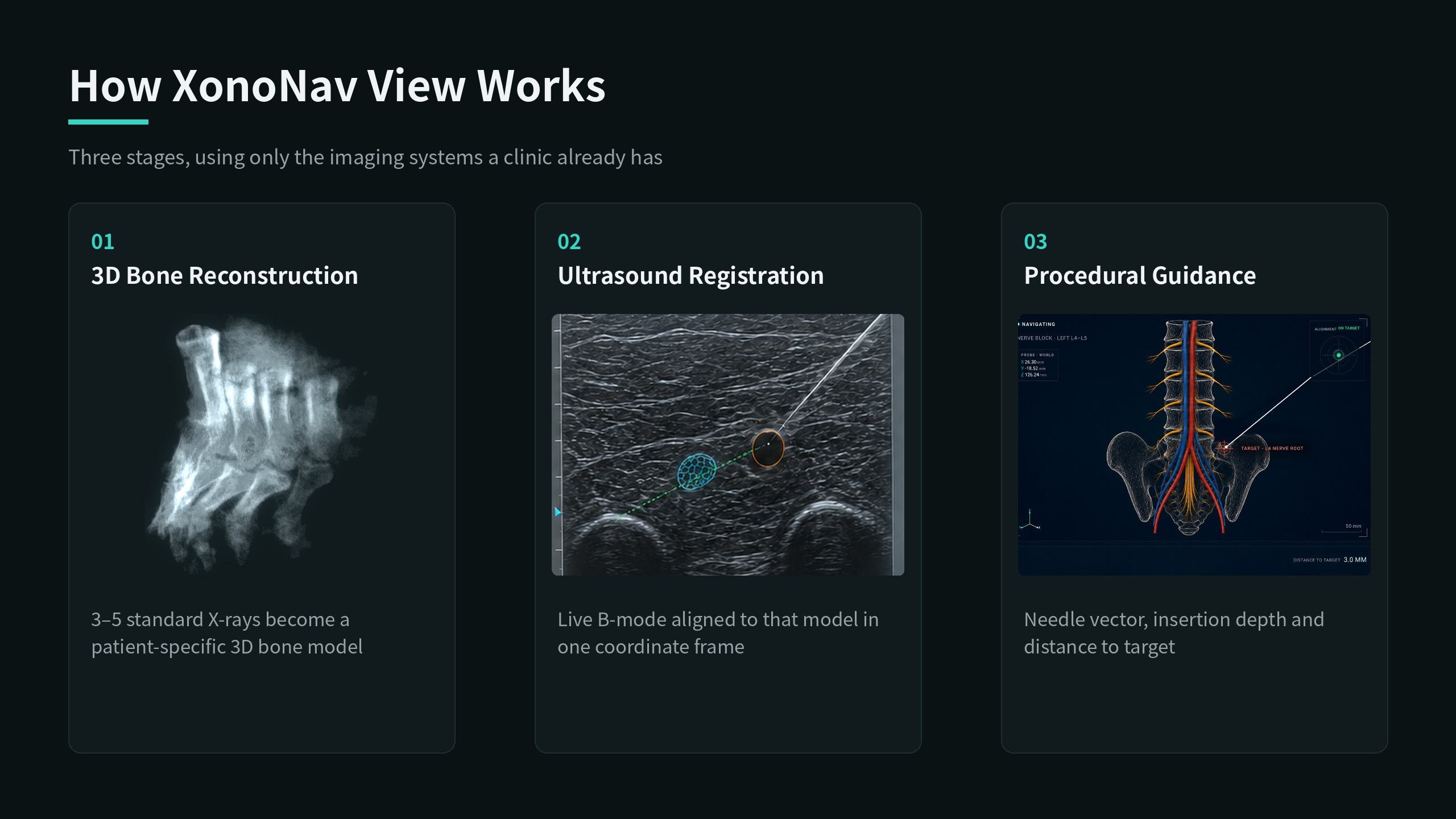
One 3D scene, built from everyday imaging
01
Patient-specific 3D from standard X-rays
Sparse-view AI rebuilds 3D bone from 3–5 standard X-rays — in under 10 seconds, no added radiation.
02
Bone and soft tissue in one view
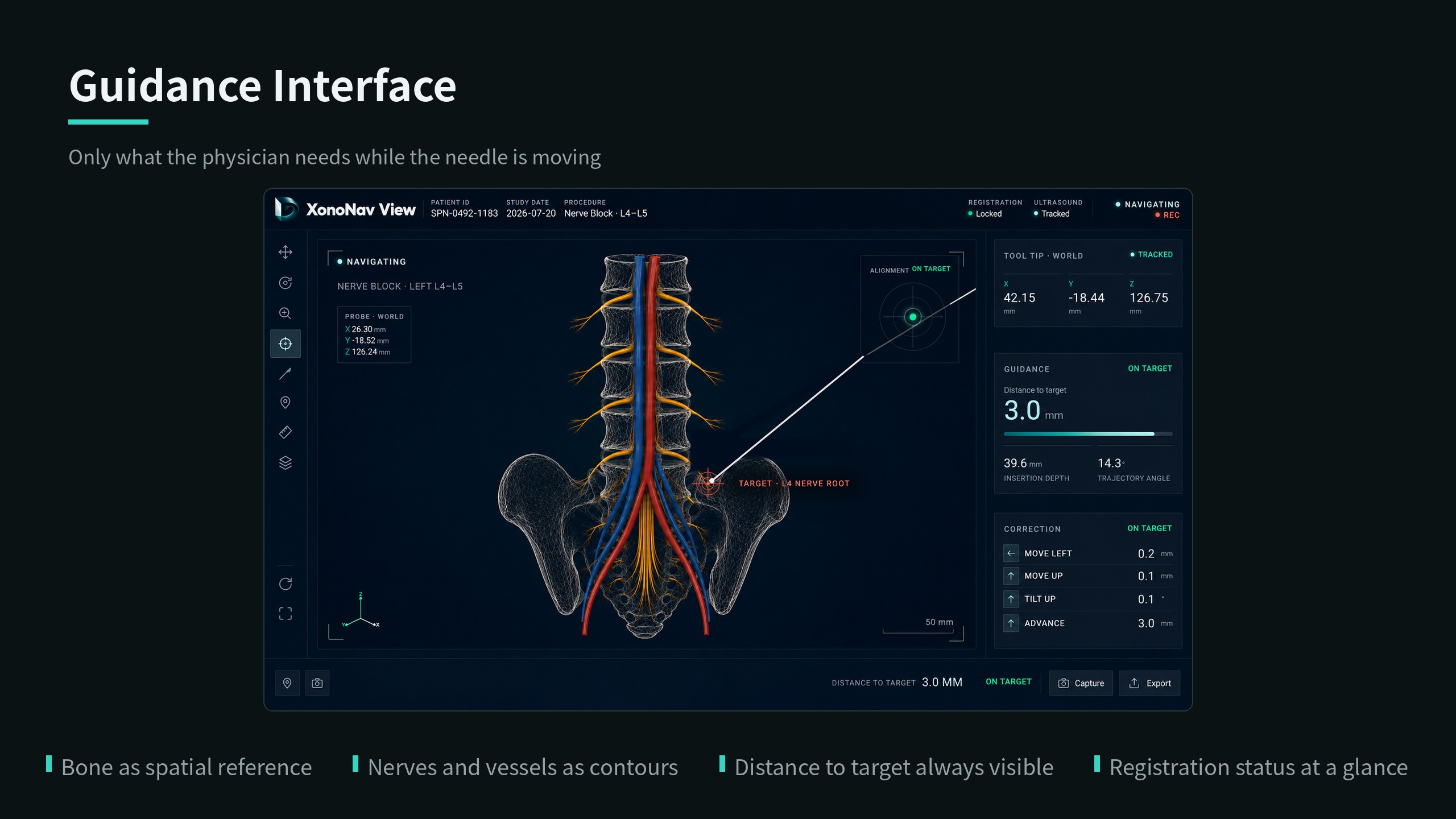
Tracked ultrasound adds soft tissue in 3D — bone, nerves, vessels, and needle in one scene.
03
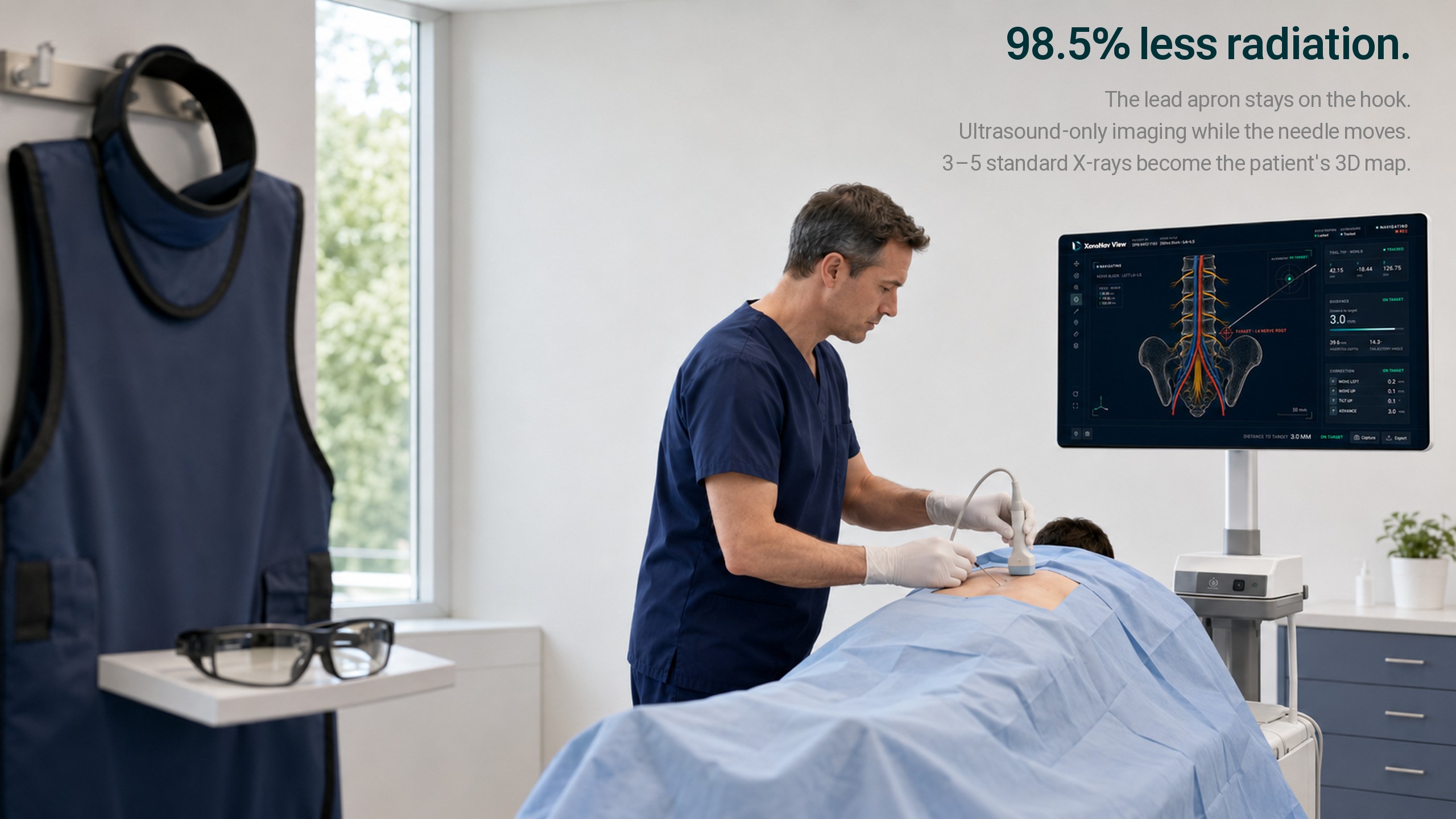
98.5% less radiation
Ultrasound-only imaging while the needle moves; 3–5 standard X-rays become the patient's 3D map — the lead apron stays on the hook.
04
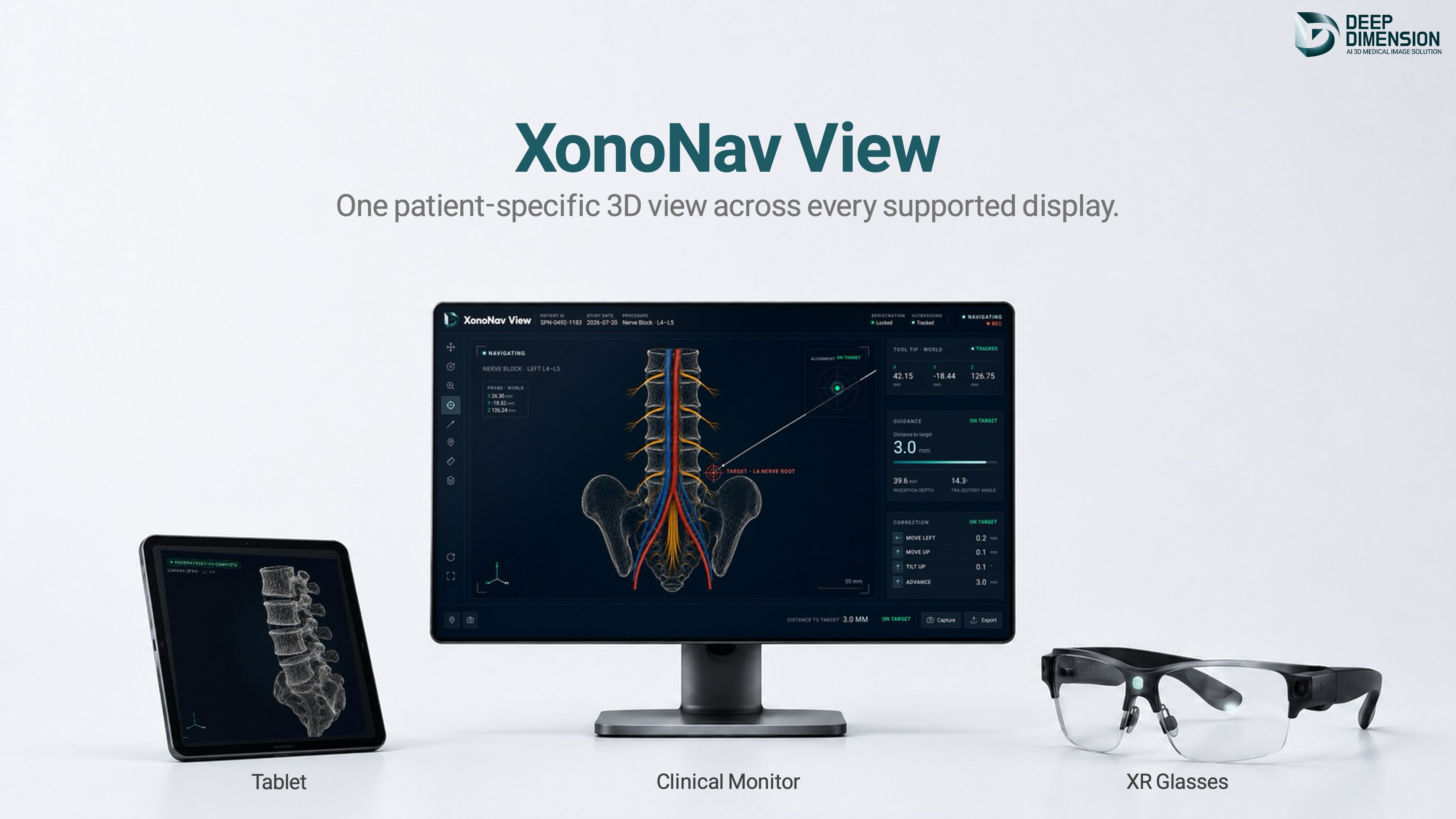
On a monitor or AR glasses
View it on a monitor or AR glasses, with an auto-generated report. Every clinical decision stays with the physician.
What it changes
Installed equipment becomes 3D navigation
No new hardware, no shielded room — the X-ray and ultrasound you already own become radiation-free 3D guidance.
standard X-ray views, already reimbursed
to build the patient-specific 3D bone model
added radiation, zero contrast agent
the 5-year cost of legacy hardware
XonoNav View is under active development. Figures reflect internal evaluation under study protocols. Not yet cleared or approved as a medical device; not offered for clinical diagnostic use.
Start with the data you already have
Your existing data is the starting point for 3D. See it yourself with a demo.